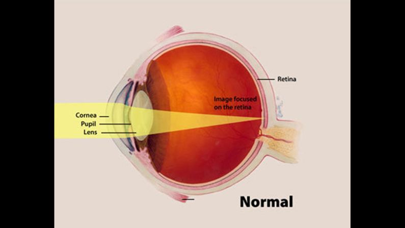
This illustration highlights the cornea, pupil and lens of a normal eye and how an image focuses on the retina.
National Eye Institute of the National Institutes of Health
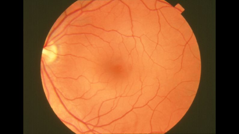
This photograph of a normal retina reveals the vascular, or circulatory, system structures.
National Eye Institute of the National Institutes of Health
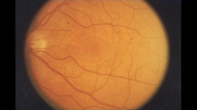
In background retinopathy, or damge to the retina, a slight deterioration in the small blood vessels occurs. Portions of the vessels may swell and leak fluid into the surrounding retinal tissue.
National Eye Institute of the National Institutes of Health
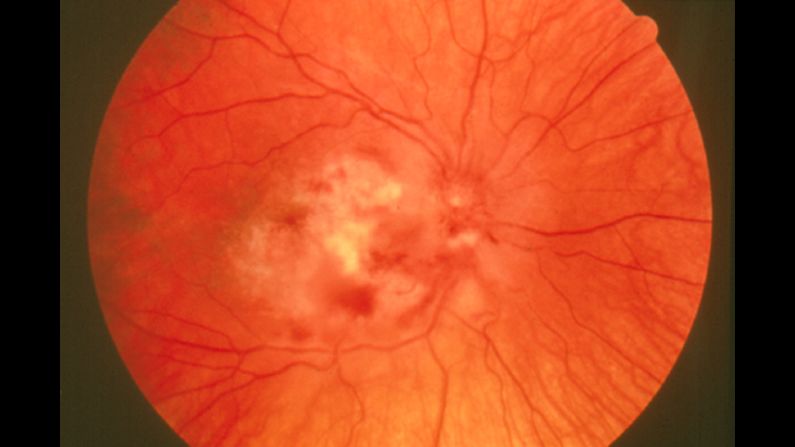
Cytomegalovirus (CMV) retinitis is caused by the herpes viruses that infects most adults. It is a serious eye infection, with symptoms that usually occur in people with weakened immune systems. Untreated, it can lead to vision problems.
National Eye Institute of the National Institutes of Health
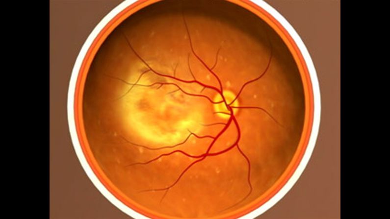
This photograph shows age-related macular degeneration, the leading cause of vision loss among people 50 and older, with fibrosis. It causes damage to the macula, a small spot near the center of the retina. The macula is the part of the eye needed for sharp, central vision.
National Eye Institute of the National Institutes of Health
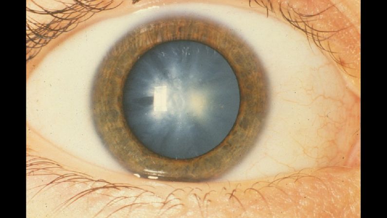
This photograph shows a white congenital cataract, an opacification (or unclear density) of the lens that was diagnosed at birth.
National Eye Institute of the National Institutes of Health
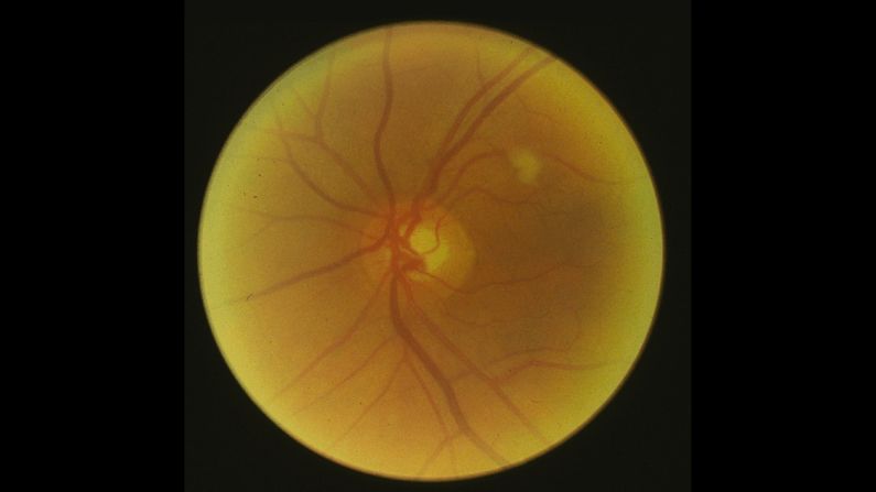
High blood pressure can cause glaucoma, a group of diseases that damage the eye's optic nerve and sometimes result in blindness. Though there is no cure, treatment in the form of eyedrops can stall the progress of this disease.
Cogan Collection, NEI/NIH
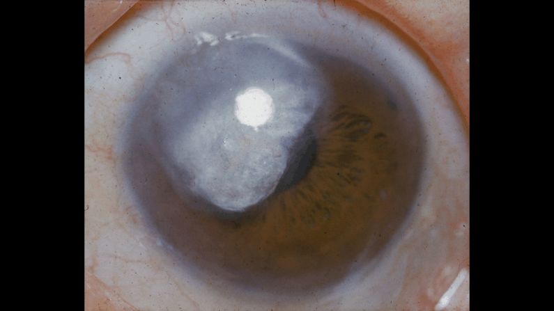
Here, glaucoma is seen, unaided by technology.
Cogan Collection, NEI/NIH
Diabetes can cause incompetent blood vessels, which leak blood into the retina. This leads to visual impairments. Diabetes affects 30.3 million US adults; one in four don't know they have it.
Cogan Collection, NEI/NIH
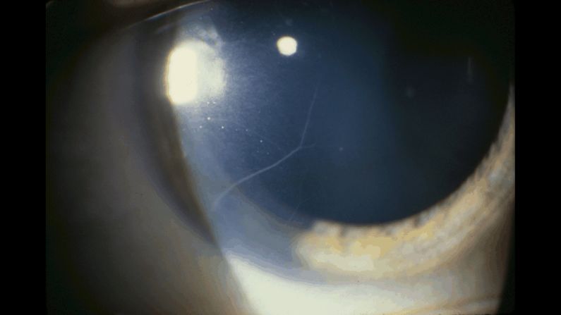
As seen in this photograph, thyroid disease may reveal prominent corneal nerves while causing dryness and bulging of the eye.