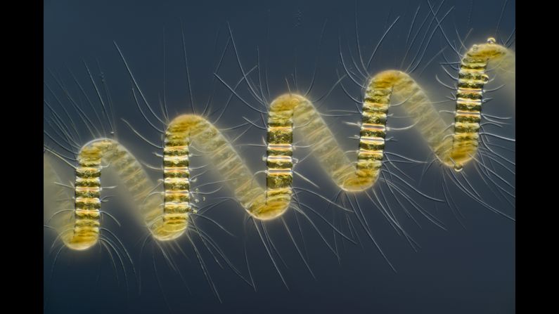
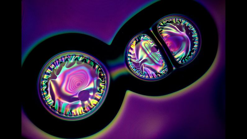
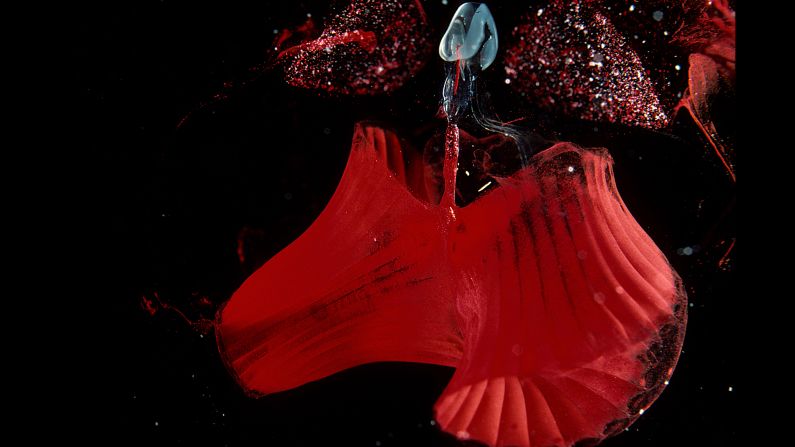
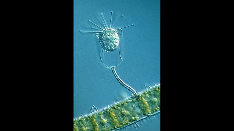
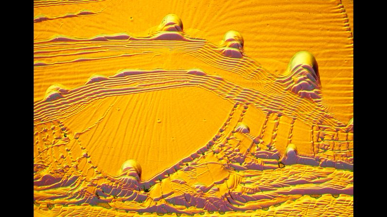
This is the 40th year that camera maker Nikon has held its Small World competition, which seeks the best magnified images melding science and art. Last year, freelance photographer Wim van Egmondwon first place for this magnified image of marine plankton. "For 20 years, I've been looking through a microscope, and every time I see things I haven't seen before," van Egmond told CNN.
courtesy Nikon Small World
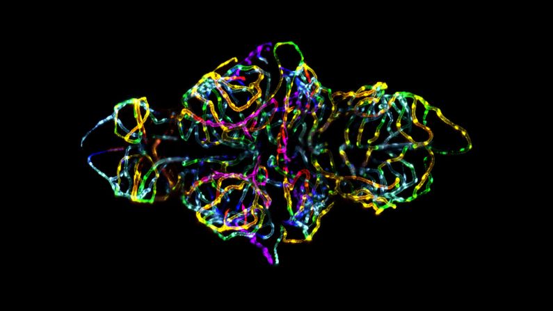
2012: Dr. Jennifer L. Peters & Dr. Michael R. Taylor, St. Jude Children's Research Hospital —
This winning image of a zebrafish embryo was injected with vibrant colors based on depth to convey spatial information, as well as to make it visually appealing. Dr. Jennifer L. Peters, one of the image's co-creators, said the photo "not only captures the beauty of nature, but is topical and biomedically relevant."
courtesy Nikon Small World
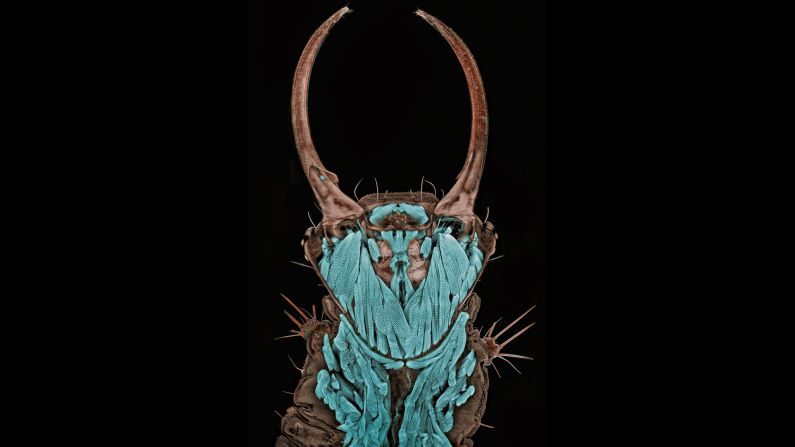
2011: Dr. Igor Siwanowicz, Max Planck Institute of Neurobiology —
"My art causes a dissonance for its viewer -- a conflict between the culturally imprinted perception of an insect as something repulsive and ugly with a newly acquired admiration of the beauty of its form," said Dr. Igor Siwanowicz, referring to his winning image of a common green lacewing. In real life, the bug's head was just 1.3 millimeters long, requiring great skill to dye and fix it in place for the shot.
courtesy Nikon Small World
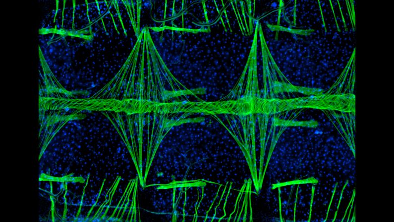
2010: Jonas King, Vanderbilt University —
This image of a mosquito heart was magnified at 100x. "Mosquitoes remain one of the greatest scourges of mankind," photographer Jonas King said in 2010. "Malaria infects hundreds of millions of people annually and is believed to have a major impact on the economies of endemic regions."
courtesy Nikon Small World
2009: Dr. Heiti Paves, Tallinn University of Technology —
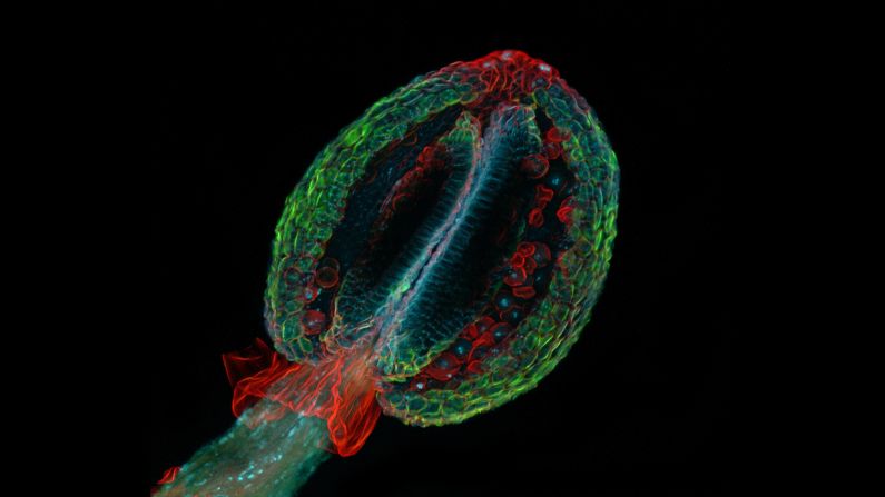
More than 2,000 entries were submitted for the prize in 2009. That year, the winning entry showed a thale cress anther, the male sex organ of a small flowering plant. "As part of my work as a research scientist, I have been taking photographs through the microscope for almost 30 years to observe the processes in living cells," researcher and photographer Heiti Paves said.
courtesy Nikon Small World
2008: Michael J. Stringer —
This exquisite image shows Pleurosigma, or marine diatoms, that were magnified 200 times. It was taken using darkfield and polarized light.
courtesy Nikon Small World
2007: Gloria Kwon, Memorial Sloan Kettering Institute —
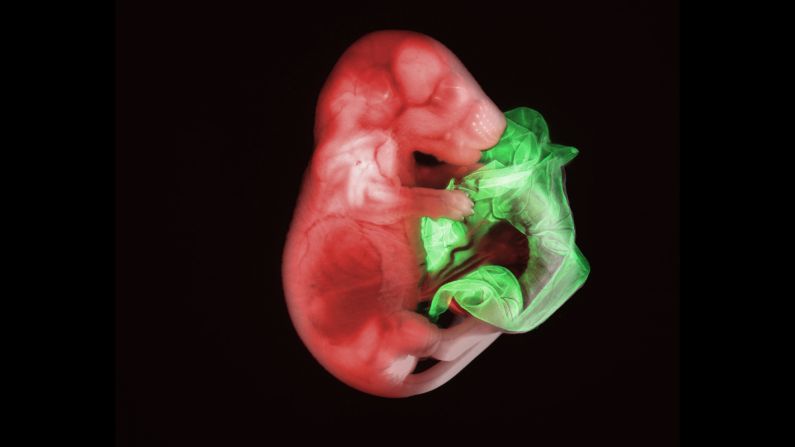
In 2007, first place went to this image of a double transgenic mouse embryo at 18.5 days. Gloria Kwon, a researcher at the Memorial Sloan Kettering Institute, used widefield microscopy and red and green fluorescence.
courtesy Nikon Small World
2006: Dr. Paul Appleton, University of Dundee, Division of Cell and Developmental Biology —
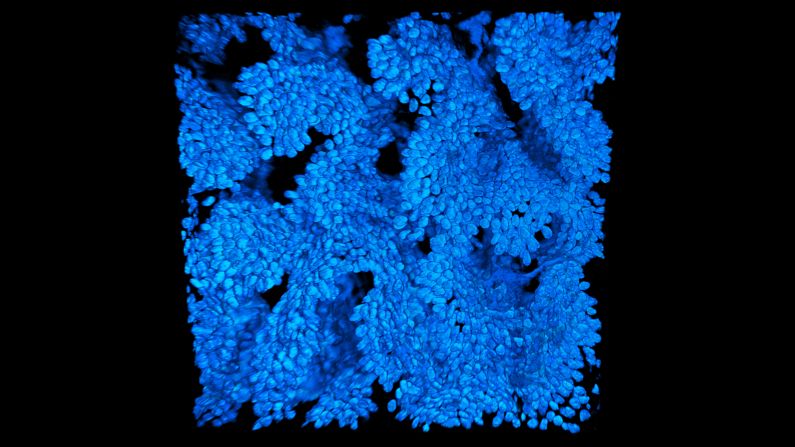
This detailed image is the cell nuclei of a mouse colon magnified 740 times. It was taken by Paul Appleton, a regular entrant to the competition who has contributed a number of "Images of Distinction."
courtesy Nikon Small World
2005: Charles Krebs —
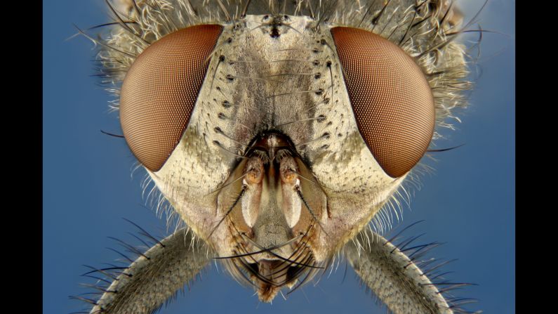
An up-close image of the common housefly took top honors in 2005. The same year, Nikon added a new award category of "Images of Distinction" to recognize some of the more notable entrants.
courtesy Nikon Small World
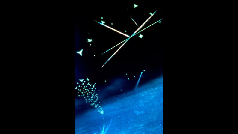
2004: Seth A. Coe-Sullivan, Massachusetts Institute of Technology —
The 2004 winner was a failed experiment, according to Seth A. Coe-Sullivan, who at the time was a graduate assistant at the Research Laboratory of Electronics at the Massachusetts Institute of Technology. The image is of quantum dot nanocrystals deposited on a silicon substrate and magnified 200 times using light reflectance. "The only reason I kept it was to document the failure. I wish I could say I spent hours getting it right, but that wasn't the case," Coe-Sullivan told Advanced Imaging Pro.
courtesy Nikon Small World
2003: Dr. Torsten Wittmann, The Scripps Research Institute —
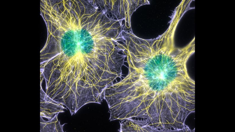
Serial place-getter Torsten Wittmann won with this shot in 2003 of magnified mouse cells.
courtesy Nikon Small World
2002: Thomas J. Deerinck, University of California, San Diego —
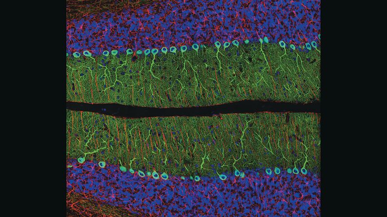
Thomas Deerinck won first prize with his first entry, a portion of a rat's brain, in 2002. He's now one of the competition's most prolific winners, and in 2008 joined the judging panel. "Aesthetics is obviously a big part of the judging, but almost as important is the technical merit of the image and how difficult or rare it was to capture," Deerinck told CNN. Capturing microscopic images requires patience, steady hands and a "lot of preparation and persistence," he said. "One of the jobs of a scientist is to convey and communicate information in a concise and accurate manner, and it helps on occasion if it also happens to be beautiful science."
courtesy Nikon Small World
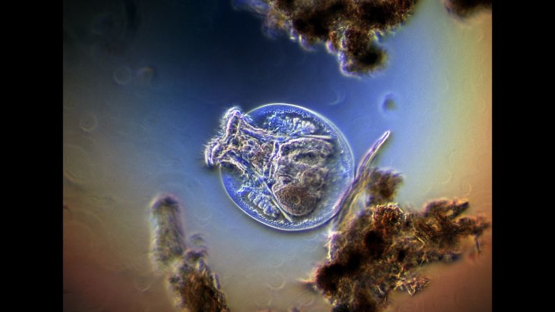
2001: Harold Taylor —
Fresh-water rotifer feeding among debris
courtesy Nikon Small World
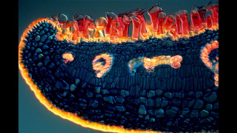
2000: Daphne Zbaeren-Colbourn —
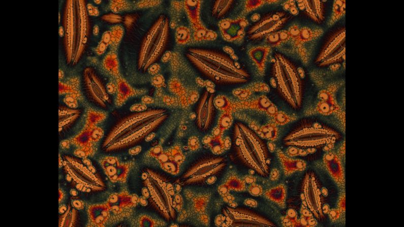
Avicennia marina (mangrove) leaf
courtesy Nikon Small World
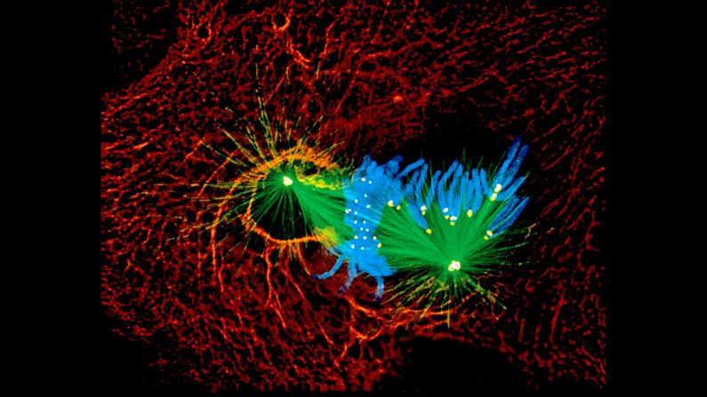
1999: Alexey Khodjakov, Wadsworth Center —
Newt lung cell in mitosis (five different structures)
courtesy Nikon Small World
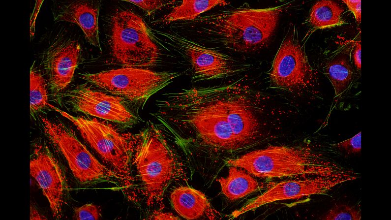
1998: Jakob Zbaeren, Insel Hospital —
Endothelial cells
courtesy Nikon Small World
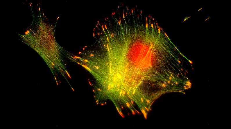
1997: Barbara Danowski, Union College —
Mouse fibroblasts
courtesy Nikon Small World
1996: Lars Bech —
Doxorubin in methanol and dimethylbenzenesulfonic acid
courtesy Nikon Small World
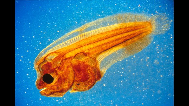
1995: Christian Gautier, JACANA Press Agency —
Larva of Pleuronectidae
courtesy Nikon Small World
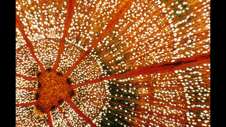
1994: Jean Rüegger-Deschenaux —
Cross section of very young beech
courtesy Nikon Small World
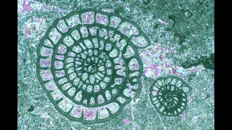
1993: Ron Sturm, Construction Technology Laboratories, Inc. —
Fossil fusulinids in limestone
courtesy Nikon Small World
1992: Lars Bech —
10-year old preparation of barbital, fenacetine, valium and acetic acid
courtesy Nikon Small World
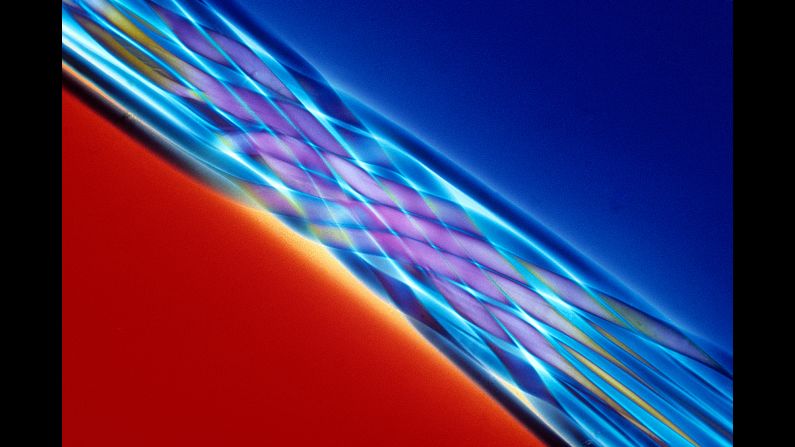
1991: Marc van Hove, Centexbel —
Polyurethane elastic fiber bundle
courtesy Nikon Small World
1990: Richard H. Lee, Argonne National Laboratory —
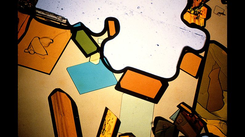
Crystals evaporated from a solution of magnesium sulfate and tartaric acid
courtesy Nikon Small World
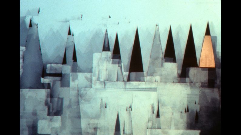
1989: Marc van Hove, Centexbel —
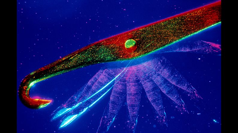
Multiple exposure of a knitting-machine needle
courtesy Nikon Small World
1988: David A. Smith —
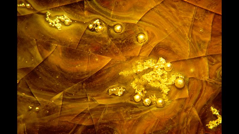
Gold residue and gold-coated bubbles in a glassy matrix
courtesy Nikon Small World
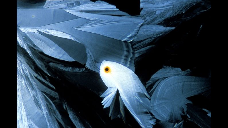
1987: Julie Macklin & Dr. Graeme Laver, Australian National University —
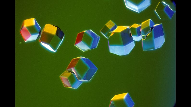
Crystals of influenza virus neuraminidase isolated from terns
courtesy Nikon Small World
1986: Dr. Stephen Lowry, University of Ulster, Department of Biology —
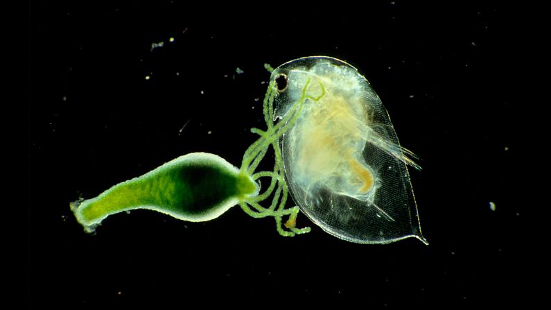
Live water mount of Hydra viridissima capturing Daphnia pulex (water flea)
courtesy Nikon Small World
1985: Dr. Jonathan Eisenback, North Carolina State University, Department of Plant Pathology —
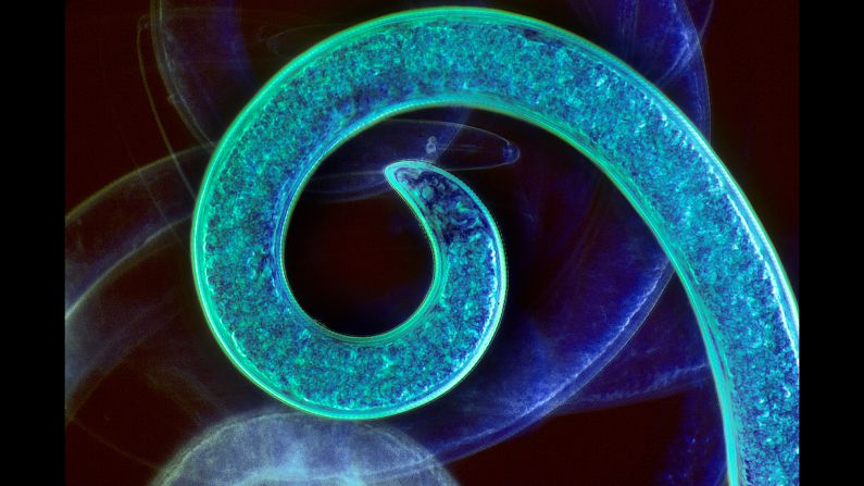
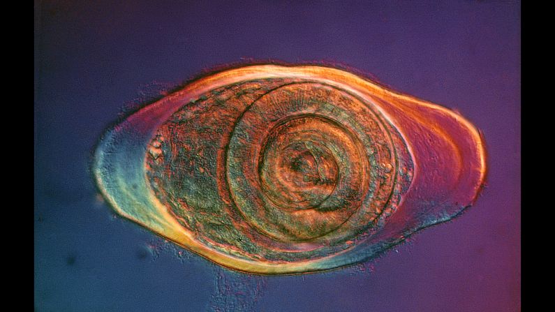
Formalin-fixed whole mount of a spiral nematode (multiple exposure)
courtesy Nikon Small World
1984: John I. Koivula, Gemological Institute of America —
Inclusions of goethite and hematite in Brazilian agate
courtesy Nikon Small World
1983: Elieen Roux, Bob Hope International Heart Research Institute —
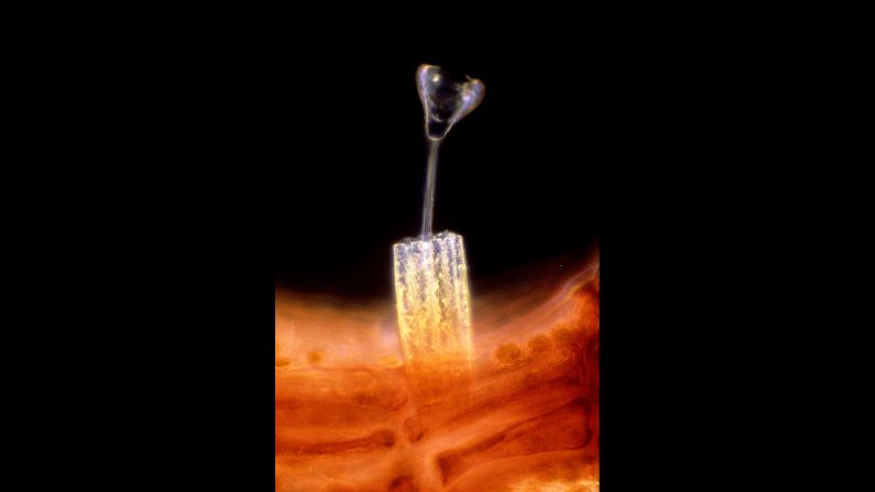
Suctorian attached to a stalk of red algae, encircled by a ring of diatoms
courtesy Nikon Small World
1982: Dr. Jonathan Eisenback, North Carolina State University, Department of Plant Pathology —
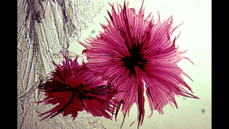
Silverberry scaly hair whole mount
courtesy Nikon Small World
1981: David Gnizak, Ferro Corporation Research Center —
Collapsed bubbles from an annealed experimental electronic sealing glass
courtesy Nikon Small World
1980: James M. King, UC Santa Barbara, Marine Science Institute —
Larvacean within its feeding structure, dyed with red organic carmine that the larvacean syphoned in while filter feeding
courtesy Nikon Small World
1979: Paul W. Johnson, University of Rhode Island —
Stalked protozoan attached to a filamentous green algae with bacteria on its surface
courtesy Nikon Small World
1978: David Gnizak —
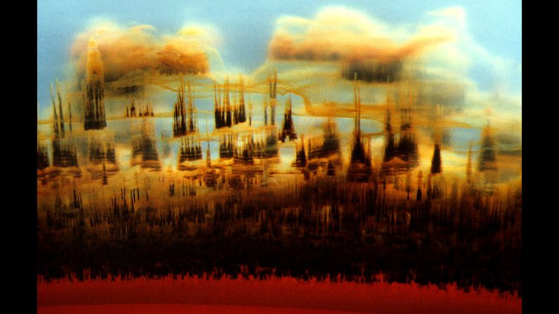
Gold, vaporized in a tungsten boat, in a vacuum evaporator
courtesy Nikon Small World
1977: James W. Smith —
Crystals of rutile (titanium dioxide) and tridymite (a polymorph of quartz) in a cobalt-rich glass